The fetus is a certain phase in the prenatal growth of viviparous organisms, which has to do with humans. The male fetus is described as the premature developmental phase of a male. The female fetus is described as the premature developmental phase of a female. Viviparous organisms possess the proficiency to put to childbearing to the live young. The live progeny is conceived inside the womb of the mommy. Immediately, the fertilization cycle occurs, and the zygote builds and grows into a fetus.
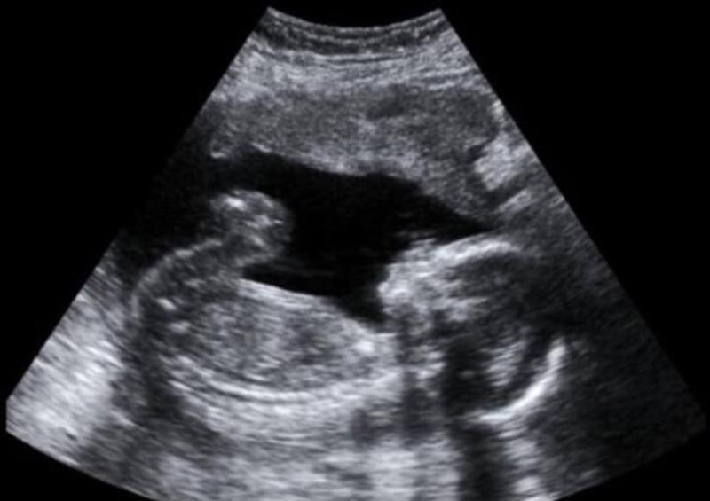
The fetus develops after the ninth week of fertilization, and it occurs between the embryonic and delivery conditions of viviparous organisms. To humans, the fetus is often observed with ultrasound scanning, with which the sex variation of the fetus could be indicated. The male fetus can be confirmed with the use of ultrasound scanning in which a lump is noticed in between the laps of the male. Verifying the female fetus with the help of an ultrasound scan reveals that there are parallel lines between the laps of the fetus which indicates the outcome of the labia and the clitoris.
The significant disparity between male and female fetuses is founded on the observance of the ultrasound scanning of the fetus. A lump is often noticed in between the laps of the male fetus. This explains the growth of the penis. A parallel line is detected in the female fetus, which indicates the development of the labia and clitoris.
What is a Male Fetus?
The male fetus is described as the premature developmental phase of the male, which is often noticed during fetus gestation. The hormonal disparity and the genetic characteristics decide the sex differentiation of a male. This is when an XY sex chromosomes duo is available in the male karyotype. The evolution of the male fetus is identified with ultrasound scanning and hence is usually indicated using various assertions. Initially, the lump in between the laps of the fetus is noticed in the ultrasound scanning of the male fetus. The growth of the umbilical cord may usually complicate this. In the second trimester, if an angle surpasses 30 degrees and gets noticed between the lump towards the left, it can be verified that the fetus is a male. Currently, fetal sex-specific biomarkers have been recognized to know the sex of a fetus. It was revealed that the male fetus indicates slow development speed in the growth of the head rim; nevertheless, at the beginning of the second trimester, they demonstrated improved head rim development. In connection to hormones, the amniotic fluid specimens often consist of more testosterone in the condition of male fetal growth.
What is a Female Fetus?
The female fetus is described as the premature developmental phase of a female, after the 9th week after it has undergone fertilization. The fetal traits of a female are relied on the parallel strings noticed between the laps of the fetus. These parallel strings match the clitoris and also the labia of a female. The unavailability of the notable lump that illustrates the male penis is also considered a trait designation facet for a female fetus. These traits are specified with the use of ultrasound scanning during the period of the first and second trimesters. The amniotic fluid specimens are high in estrogen at the time of the female fetal growth when comparing the female fetus with the male fetus. The fetal sex differentiation is as well attributed to various biomarkers like the growth of the head rim, crown posterior height, and femur heights. It was noticed that the female fetus indicates an increased crown chunk length when comparing it to the male fetus, while the attributes, which include the head rim and femur height, are lower in a female fetus.
Difference Between Male and Female Fetus
The male fetus is described as the premature developmental phase of a male. The female fetus is described as the premature developmental phase of a female. The male fetus is often verified with ultrasound scanning, in which the lump is noticed between the laps of a male. The female fetus is confirmed with ultrasound scanning, indicating that there are parallel strings between the fetus’s laps. This signifies the growth of the labia and the clitoris. There are increased levels of testosterone and reduced estrogen levels in the male fetus. The female fetus includes increased estrogen and decreased testosterone level.