Teratoma and seminoma are germ cell tumors with comparable features but vary in pattern. Teratoma is a properly encapsulated tumor possessing elements obtained from every three germ coatings, while seminoma originates from the germ cell epithelium of the seminiferous tubules. This article highlights the difference between these two words.
What is a Hematoma?
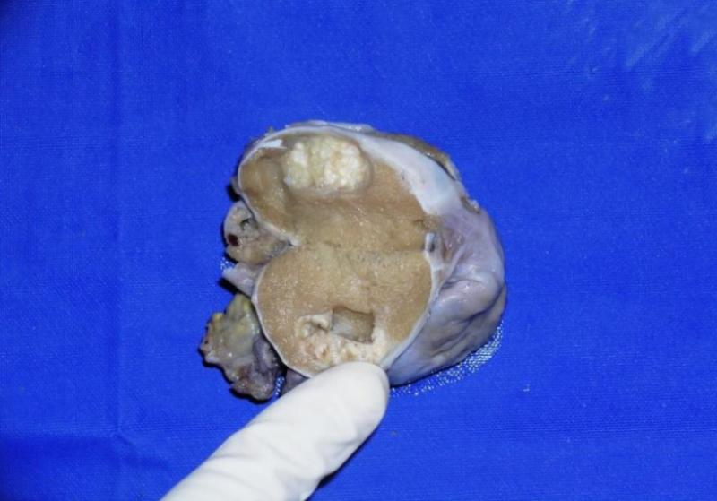
As earlier stated, it is an adequately encapsulated tumor possessing elements acquired from every three germ cell coatings. It is categorized as ripened and premature, in which the latter is malignant. Every teratoma in older individuals is naturally malignant; conversely, in children below 12 years, they act as a benign neoplasm. As it is regarded to be a congenital tumor, it exhibits at birth. But a lot of times, the tumors are signaled at a mature age. Alpha-fetoprotein can be enormously increased. Microscopically, teratoma displays somatic distinctions and consists of a component of every three germ coatings: mesoderm, ectoderm, and endoderm. It may have to do with brain hair, skin, teeth, respiratory and intestinal mucosa, bone, and cartilage. When it has to do with a fetus, they are not harmful but can hardly trigger massive impacts and vascular stealing, which may result in fetal heart delinquency. Treatment has to do with total surgical excision of the tumor. For malignant teratomas, chemotherapy is provided instantly after the surgical operations.
What is Seminoma?
It is the vastly treatable and fixable cancer found in the testis. It often emanates from the germinal epithelium of the seminiferous tubules. Half of the germ cell tumors found in the testis have to do with seminoma. If it takes place in the ovary, it is described as dysgerminoma, whereas in the central nervous structure, it is described as germinoma. Clinically, the individual shows up with a testicular mass, back pain, testicular pain, and testicular atrophy, ensuing metastasis to the vertebra. Analysis research concerns elevated levels of alkaline phosphatase and increased human chorionic gonadotropin. When it involves the masterpiece seminoma, serum alpha-fetoprotein is not increased. Macroscopically, it displays as a meaty and lobulated gathering. The tumor protrudes from the cut veneer, and hemorrhagic regions may be noticed. Using a microscope, a masterpiece seminoma can be featured by perch of livery enormous round cells which possesses different cell membrane, well-known nucleoli, clear cytoplasm, and central nucleic which consists of increased glycogen, which looks like the main spermatocytes in the seminiferous tubule. Other types have to do with spermatic seminoma featured by the development of the tumor cells, which corresponds to secondary spermatocytes. Ana plastic seminoma is extremely pleomorphic and has increased mitotic sculptures. Treatment has to do with inguinal orchidectomy in nearly every possibility. The tumor also displays a dramatic keenness to radiotherapy and chemotherapy, with a reasonable survival status of about 90 percent in the beginning phases.
Difference Between Teratoma and Seminoma
- Teratoma is a properly encapsulated tumor possessing elements acquired from all three germ coatings, whereas seminoma is obtained from the germinal epithelium of the seminiferous tubules.
- Teratoma is properly encapsulated.
- Regarding teratoma, the difference between benign and malignant is in the collaborative development of the patient’s member locations, tissues, and age. In contrast, seminoma is a vastly treatable and fixable cancer in the beginning phase.
- Heightened alpha-fetoprotein stages are generally connected with teratoma.
- Treatment of teratoma involves total surgical excision of the tumor, whereas, in seminoma, inguinal orchidectomy is required in nearly all conditions.